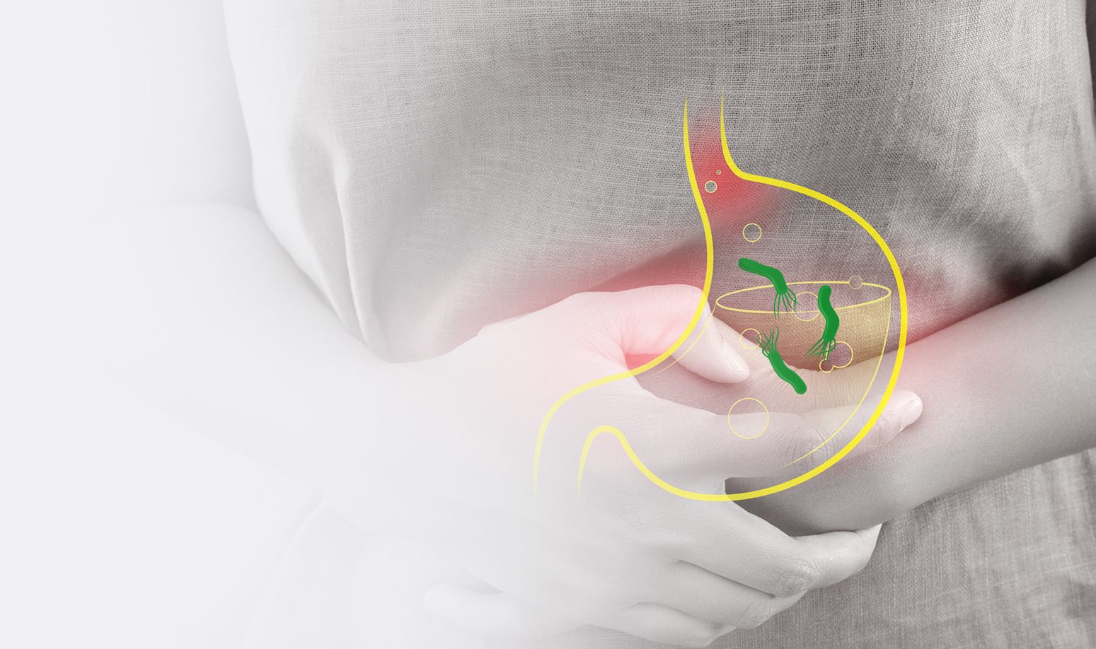
Advantages of Quantum Dot technology by comparison with the existing diagnostic methods for Helicobacter pylori.

Diagnosis of Helicobacter pylori has so far been based on two groups of tests: invasive diagnostic tests, which include endoscopy, histological analysis, rapid urease test and bacterial isolation, and non-invasive diagnostic tests, which include urea breath test, H. pylori stool antigen test and serology tests for H.pylori. 

helicobacter 1

Non-invasive diagnostic tests:

a) Urea breath test: based on the fact that H.pylori produces urease, which catalyses the hydrolysis of urea to ammonia and carbon dioxide. The patient drinks the solution of urea with 13C or 14C carbon isotopes, which is then disintegrated by urease should H.pylori be present. The released 13CO2 or 14CO2 first gets into the bloodstream, then into the lungs, where they are measured by the breath test 30 minutes after the intake of urea in the body. The test is highly specific and sensitive, yet false positive results may occur in the presence of other microorganisms that produce urease in the human stomach. In patients treated by proton pump inhibitors as well as those infected by a metabolically inactive coccoid form of H.pylori, the results may be false negative, and test sensitivity is also lower in patients who have undergone stomach surgery. Also, the 14C carbon isotope is not recommended for use by children and pregnant women due to its radioactivity.

b) H. Pylori stool antigen testing: there are two types of antigen tests available in diagnosing H.pylori infection-enzyme immunoassay based on monoclonal antibodies and the immunochromatographic test. The accuracy of this test’s results may be diminished by the patient’s failure to stop using the proton pump inhibitor at least 4 weeks prior to the testing, due to the fact that the stool sample is inhomogeneous, so the sample may be taken from a place with no H.pylori. Furthermore, a watery stool sample as well as the presence of bleeding in the upper part of the gastrointestinal system may also affect the accuracy of results

c) Serological tests: most of the time patients have a chronic infection caused by H.pylori, and as such IgG antibody tests need to be used. The most commonly used tests include ELISA, immunochromatography, and Western blot. Antibodies may remain in the body even years after healing, which is why serological tests cannot be used for estimating the success of healing.

Invasive diagnostic tests:

Invasive diagnostic tests are commonly used with patients over 50 and when alarming symptoms occur regardless of the patient’s age.

a) Endoscopy: used to visualise the entire upper part of the gastrointestinal tract (esophagus, entrance to the stomach, corpus and its exit as well as the beginning of duodenum), which, on one hand, causes discomfort in the patient, while on the other hand, the use of the endoscopic procedure alone, without additional diagnostic tests, does not allow diagnosing the presence of H.pylori as there is no “typical sign” of its presence.

b) Rapid urease test: tests the presence of H. pylori in the sample obtained by the biopsy of gastric antrum. As the test is based on enzymatic reaction, parameters that affect the reaction, such as substrate concentration, concentration and activity of the enzyme, temperature and time, should be considered. For a positive urease test the biopsy sample should contain a minimum of 100 000 H.pylori bacteria in order for the indicator colour to change. Also, the sample should be taken from the exact spot where H.pylori is located. The test result may also be affected by medication (H2 receptor antagonists, medicines containing bismuth, proton pump inhibitors, antibiotics). It is important to note false positive results of the test, which may occur at the release or urease from other bacteria that produce it, such as Klebsiella pneumoniae, Staphylococcus aureus, Proteus mirabilis and Enterobacter cloacae. Sensitivity and specificity of rapid urease tests is also lower in patients with acute gastrointestinal bleeding.

c) Histological analysis of the stomach biopsy sample:represents a highly sensitive method, however, there are several factors that impede the diagnostic accuracy of the method. They include the place and the number of taken samples (for a complete histological analysis as many as five samples are required, two from the antrum, two from the corpus and one from the angular part), choosing a colouring method, former medical treatments and the experience of the pathologist who analyses the specimen.

d) Isolation of H.pylori from the biopsy sample: the method has a high specificity level of nearly 100% and a level of sensitivity that varies between 85% and 95%. However, poor sample quality, slow transport, exposure to an aerobic environment, highly active gastritis, alcohol consumption and long-term use of histamine receptor antagonists may have a negative impact on the accuracy of this diagnostic method.

In recent times, molecular diagnostic methods – RAPD-PCR (randomly amplified polymorphic DNA), RFLP-PCR (restriction fragment length polymorphism) and DNA fingerprinting–have been available for H.pylory diagnosis and typification. However, these methods are not widely used outside major hospitals.

Quantum Dot method:

It is the possibilities offered by molecular diagnostics, as well as the wide accessibility, shortness of time required to obtain results, and a high level of sensitivity and specificity in H.pylori diagnosis and typification, that prompted Chronolab to introduce Quantum Dot technology. It represents a revolutionary immunochemical method as it represents a “double sandwich method” used for detecting antibodies to urease, CagA and VacA antigens.

A reagent represents an antibody that reacts to the serum antibody, and in this method, the antibody is the detected antigen. During the reaction of antibodies and antigens (antibodies), a signal is created which the analyser converts into a numerical value of the measured antibodies.

Urease makes it easier for H.pylori bacteria to survive in the stomach’s acid conditions, whereas the most important H. pylori virulence factors are the vacuolating cytotoxin A (VacA) and the cytotoxin-associated geneA (CagA). Upon endocytosis, VacA forms vacuoles thus damaging endothelial cells. CagA belongs to the cag pathogenicity island (cagPAI), which consists of 31 genes encoding T4SS. T4SS is a needle-like structure that penetrates through the membrane of gastric epithelial cells inserting H. Pylori products including CagA. CagA is the most virulent factor related to this bacterium.

The use of the Quantum Dot method allows:

  • Avoiding testing discomfort (testing is carried out on a blood sample)
  • Wide testing availability 
  • Avoiding negative impacts of sample transport on the result 
  • Simultaneous detection of antibodies to three pathogenicity factors of H.pylori
  • H.pylori typification and determination of the degree of its pathogenicity depending on the positivity of certain antibodies (CagA and VacA antibody positivity leads to the development of stomach ulcers and cancer)
  • Full automation of the process with a quantitative presentation of results
  • Avoiding the impact of the current medical treatment on test results
  • Avoiding the impact of food and alcohol on test results
  • Avoiding the impact of possible bleeding in the gastrointestinal system
  • Avoiding false positive results due to the presence of other bacteria producing urease
  • The possibility of testing children and pregnant women because there is no radiation (as with the use of carbon 14C in the breath test)
  • Estimating the need for eradication therapy
  • Adequate monitoring of the success of the eradication therapy

References:

  1. Vaira D, Holton J, Menegatti M i sur. Review article: invasive and noninvasive tests for Helicobacter pylori infection. Aliment Pharmacol Ther 2000;14(Suppl 3):13–22. 

  2. Marušić M, Presečki V, Katičić M, Dominis M, Kalenić S. The place and role of serologic methods in detecting Helicobacter pylori infection. Coll Antropol 2006;30:529–33. 

  3. Yakoob J, Jafri W, Abid S i sur. Role of rapid urease test and histopathology in the diagnosis of Helicobacter pylori infection in a developing country. BMC Gastroenterol 2005;1:38–42. 

  4. Stolte M, Meining A. The updated Sydney system: classification and grading of gastritis as the basis of diagnosis and treatment. Can J Gastroenterol 2001;9:591–8.